Blood Smears
23.6 Blood SmearsNote: The change to pitch (12) and font (6) must be converted manually.
Making a blood smear is the process of taking a drop of blood and “smearing” it out in a thin layer on a microscope slide. For diseases like Babesiosis and Trypanosomiasis, it is very important to take a blood smear and actually see the organism. Even if a microscope is not available, prepare a blood smear and send it into a laboratory for examination.
How to Make a Blood Smear:
For Babesiosis and Trypanosomiasis, a fairly thick smear of blood is needed. For Anthrax and other diseases, a thin blood smear is best.
For live animals: Only a tiny drop of blood is needed. Therefore, it is easiest to take a sample from an ear vein for a blood smear.
Identify an ear vein and puncture it with a sharp, sterile needle.
For dead animals: Make a blood smear of blood from the ear. Be careful to wear gloves, and take only a tiny drop if anthrax is suspected.
Thin Blood Smears for Anthrax and Other Diseases:
Remember - be careful if you suspect anthrax. A tiny drop of blood from the ear of the dead animal is enough. Protect your hands.
A very small drop of blood is placed near the end of a clean glass slide.
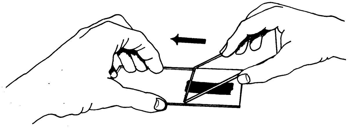


Quickly, another clean glass slide is taken and the drop of blood on the first slide is touched with an end of the second slide. This allows the blood to spread evenly along the edge.
The top slide is held at an angle of about 45 degrees to the bottom slide which should be on a flat, level surface.
The top slide is pushed gently and smoothly along to the end of the second slide, drawing the blood along behind it.
The smear is then allowed to dry. A second smear should also be made.
Do not make the smear too thick or it will be difficult to examine.
After the second smear is made, the two slides are placed together, back to back, with the blood surfaces outward. They are wrapped in a thin piece of paper.
Thick Blood Smears e.g. for Babesiosis and Trypanosomiasis:
A small drop of blood is placed near the center of a clean glass slide.
Spread out the blood with a clean match stick (Rinse the match stick ahead of time and allow it to dry).
Do not make it too thick or it cannot be examined.
The blood is then allowed to dry.
A second smear is also made and allowed to dry.
After the second smear is made, the two slides are placed together, back to back, with the blood surfaces outward. They are wrapped in a thin piece of paper.
These samples, along with the report which describes the case, are sent into the lab.