Lameness : Fractures, Broken Bones, Hoof Problems, Wounds, Arthritis, Nutritional Deficiencies
15.2 Lameness Lameness is usually due to:
1. Injury to:
-Bone, See page 233.
-Muscle, See page 209.
-Hoof, See page 236.
-Skin, See pages 235-236.
-Nerve, See pages 209, 259.
2. Bone or joint infection, See page 234.
3. Mineral deficiency, See page 242.
4. Inflammation of the joints, See page 238.
57 Sometimes an animal cannot walk properly. The term for this general condition is “lameness.” If an animal cannot move at all, the condition is called “paralysis.”
Lameness can be acute or chronic. In either case one must always check first for a fracture.
Lameness with Fractures A fracture is a broken bone. A fracture is often accompanied by injury to the surrounding tissues, including blood vessels and nerves. Fractures can result from falling, colliding with something, or being hit by something.
Fractures can be divided into three types:


1. Simple fracture: The bone is broken in only one place, and there is no wound in the skin.
2. Compound fracture: The bone is broken in more than one place, but there is no wound in the skin.
3. Open fracture: A wound in the skin, resulting in the bone being exposed, accompanies the fracture (either simple or compound) and infection may result.
Examination of Fractures: When a fracture is suspected, handle the animal carefully to avoid further injury to the animal or to the people handling it. Sometimes, the animal may be very frightened or stressed by the pain and won’t become calm until the fracture is stabilized. Examine the fracture thoroughly. Check for wounds, bleeding and whether a joint may be involved in the fracture. Examine the entire animal for other injuries.
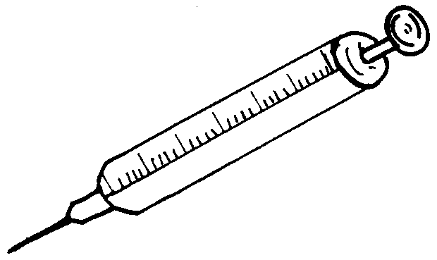
Some fractures are difficult to diagnose without the aid of an x-ray. An x-ray is a picture of the bones taken by a special machine. However, even without an X-ray machine, the following signs can help to diagnosis fractures:


Severe pain or local tenderness, resulting in the animal limping, not bearing weight on the affected limb, not standing up, or reacting strongly when the affected area is manipulated.
Deformity or abnormal angle of the bone.
Local swelling.
Crackling noise when the affected area is manipulated - due to the broken bone ends rubbing against each other.
General Treatment of Fractures
Most fractures will heal if the fractured ends are placed close to each other and immobilized. However, infection or involvement of a joint may complicate healing. The following steps should be taken to treat a fracture:
1. “Clean the wound” asso-ciated with the fracture.
2. “Set” the broken bones.
3. “Fix” the broken bones.
4. “Provide supportive care” for the animal until it recovers.
58

1. “Clean the wound” (if there is one). Open fractures require special care to avoid serious bone infections. The wound must be thoroughly washed and cleaned. Antibiotic cream should be applied topically. The wound should be protected from flies. Penicillin injections should be given to prevent infection.
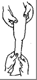
1. “Set” the bones: In any fracture, the broken ends of the bones should be adjusted so that they are properly aligned, and as close as possible to each other. This is called “setting” the fracture. Sometimes setting the bones is difficult because the muscles contract and pull the bones out of alignment. Several strong people may be required to align the bones.


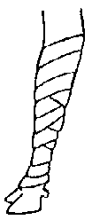


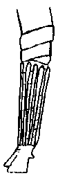


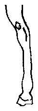
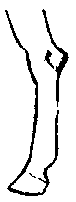
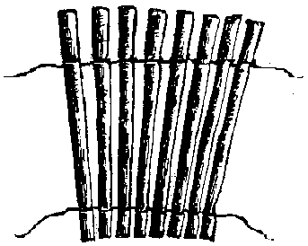
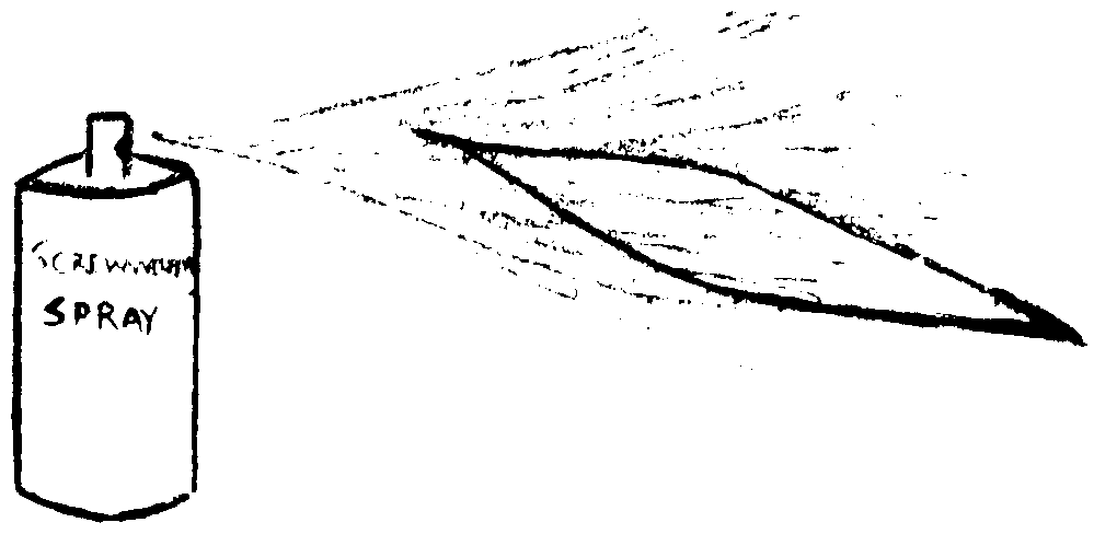
3. “Fix” the bones: Once the bones are set, they must be “immobilized” (i.e. held in place so that they do not move while healing takes place). This is called “fixation”, or “fixing the fracture.” There are many different ways to “fix a fracture” so that the bones cannot move. One easy method is to use splints made from strips of wood (like bamboo). Wood splints are less expensive, more easily available, and more lightweight than plaster. They can be easily made, but do get ruined when wet (like plaster does). Wood splints can also be removed easily and replaced to check and treat wounds associated with the fracture. A bandage should be applied under the splint for padding, and extend beyond the edges of the splint. The bandage should be sprayed or sprinkled with insecticide powder to prevent maggots/screwworms, particularly if there is a wound.
Splint made from Apply a bandage for padding Apply the splint, including the bamboo or wood. to prevent sores. Insecticide joint, above and below the should be applied to the bandage fracture. The bandage under the beforehand to prevent maggots splint should extend beyond the or screwworm. splint.
4. “Provide supportive care for the patient”: See page 234. This means regularly checking the splint, turning the patient over periodically, providing high quality food, clean drinking water, and adequate shelter away from other animals that might cause more injury or stress.
Supportive Care for a Fracture Patient:
See page 78 for general information on First Aid. In addition, the following are guidelines that apply specifically to fracture patients.
1. Time required: In general, a splint should remain on small animals for at least one month, and on large, heavy animals for at least two months. However, the time required may vary considerably depending upon the following factors:
Age of the animal: Young animals heal faster than old animals.
Size of the animal: Small animals heal faster than large animals. Large animals may sustain permanent muscle damage if they cannot stand up and are lying down for longer periods on a hard surface without moving.
Temperament of the animal: It is more difficult to properly treat and care for an excitable animal with a fracture than one that is easy to handle.
Type of fracture: Simple fractures heal faster than open or compound fractures.
Location of the fracture: Fracture of large bones, or high on a leg take longer to heal than small bones, or fractures further down. Fractures that involve a joint may take longer to heal, and the animal may develop arthritis due to permanent joint damage.
Ability to properly set the fracture: Fractures that are poorly aligned may take longer to heal.
Ability to immobilize the bone: Fractures that are not properly immobilized will take longer to heal.
Blood supply: Fractures that have a good blood supply heal more rapidly than those without a good supply.
Infection: An infected bone, soft tissue, or joint will delay healing, or even prevent it.
Consideration should be given to all of these factors before treating a fracture. Several factors together may considerably increase the time for healing or even prevent healing. In this case, the owner may prefer to slaughter the animal.
Keep the animal on soft ground and turn the animal over from side to side several times daily: This prevents them from getting any wounds on their skin from laying in the same position. It also helps blood flow to the fractures which encourages faster healing. The animal (especially big, heavy ones) should be kept on soft ground particularly if it cannot stand up. Otherwise, the pressure of its own body may cause permanent muscle damage.
1. Lift the animal: If possible, the animal should be lifted to its feet at least once per day. The animal can then support itself for a while on its other legs. This will help the animal’s blood circulation and muscles.
1. Provide good food, water, shelter and protection from other animals: Animals that are healing from a fracture need high quality food, clean, fresh water, and good shelter from heat, excessive sun, or cold. They also need protection from other animals that might injure it.
1. Watch/smell for maggot infestations, wound infections or dying tissue: Wounds should be observed daily for possible maggot infestations or wound infections. As a preventive measure, the bandaging and wound itself (if there is one) should be sprayed or dusted with insecticide powder or liquid that is effective against maggots/screwworm. The bandage and splint should be applied correctly to avoid circulation cut-off. Animals with open fractures should receive antibiotic injections such as penicillin.
Nevertheless, if the animal suddenly stops eating, has a fever, smells bad (i.e. rotten smell coming from the area of the fracture) or seems to be in unusual pain, the splint should be removed immediately. Then examine the fracture for possible infection, maggot/screwworm infestation, sores, or poor blood circulation. Poor blood circulation may result from the splint or bandage being too tight, or pressing against an important blood vessel. It also may result from not enough padding under the splint. Sometimes the arteries which supply blood to the bone and surrounding tissues are permanently damaged by the same accident which broke the bone. If the arteries are permanently damaged, the fracture may never heal.
Dislocation of Joints Sometimes the bone itself is not broken but instead the bones come out of their proper position within a joint. This is called a “dislocation.” If the muscles and tendons which hold the joint in place have also been torn (i.e. not just stretched), then the condition may be serious.
Treatment of dislocations: First, try to push and pull the joint back into the correct position. (If there is a grating sound when moving the bones, it means there is a fracture also.) Then treat the animal like a fracture patient:
1. For joints in the upper leg, simply rest the animal and give it proper daily care.
2. For joints in the lower leg, hold the bones in place with the use of a splint.
3. Provide good supportive care as for a fracture patient.


Lameness without Obvious Fractures Lameness due to Wounds:


Lameness may be due to wounds almost anywhere on the leg. Depending on the nature of the wound, it might be possible to suture it. See pages 213-214.
Treatment: All wounds should be cleaned thoroughly, and any maggots or screwworms should be removed. Ointment or spray should be applied to prevent maggots or screwworms.

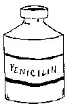


If the wound is infected, then an antibiotic injection should also be given (particularly if there is a risk of tetanus).
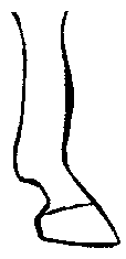
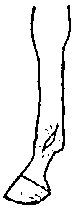
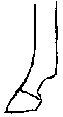
Lameness due to Wounds or Abscesses around the Hoof One of the most common causes of lameness is a wound or abscess in the hoof or near the hoof. (The hoof is part of the Skin System but it is included here with other lameness problems.)
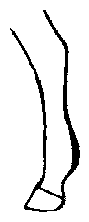
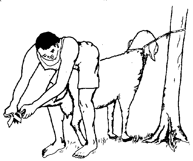
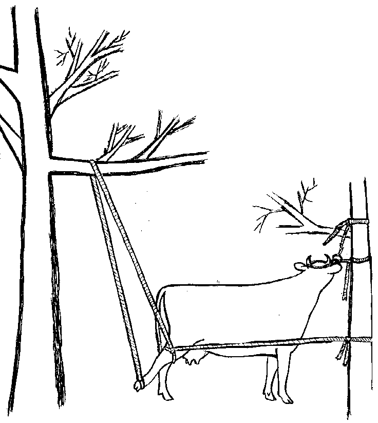
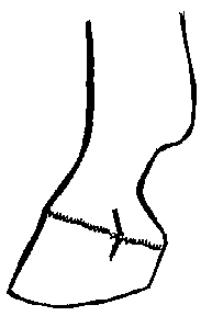
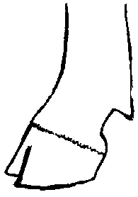
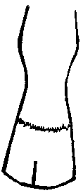
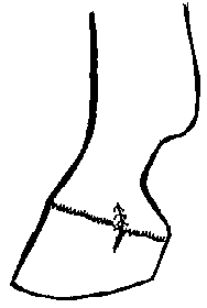
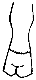
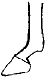
Examination of Hooves The hooves of horses, goats, and sheep can often be examined while the animal stands, by lifting the foot off the ground. For cattle/buffalo, each leg can be hoisted with a rope, or the animal should be cast.
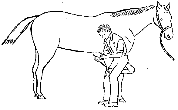


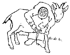


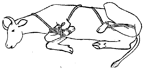
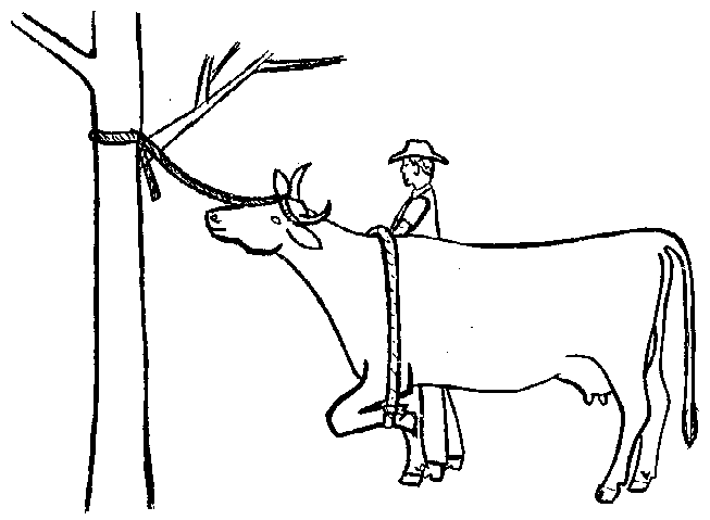
After the animal is cast, the hoof is cleaned thoroughly and pressed in various places to see if any certain spot is sore (i.e. upon pressing a sore spot, the animal will quickly withdraw its leg).


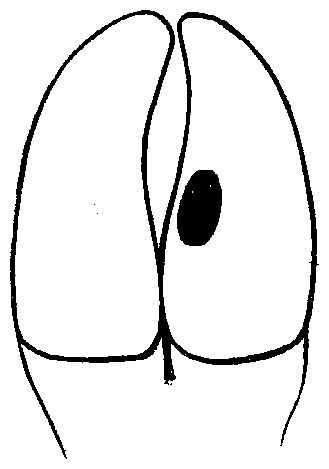
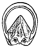
Next, look for small dark spots on the bottom side of the hoof which may indicate an abscess or a bruise, particularly at any spots that are sore. These spots should be carefully trimmed with a hoof knife until they are open and shallow. If pus comes out, it is an abscess. If blood comes out, it is a bruise.
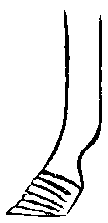
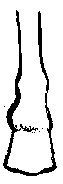
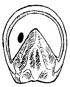
Horse hoof Cow, goat, sheep, llama, alpaca hoof


On the bottom-side of a horse’s hoof, push on the soft, triangular part called the “frog.” If pus comes out, there is an infection within the frog. See next page for details of treatment. Keep the animal in a dry, clean area.


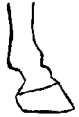
In animals with a cloven hoof, the space between the toes should be carefully examined for any wounds due to a foreign object (e.g. a stick).
The coronary band should be examined for any wounds.


The entire hoof should be examined for cracks or splits. If severe enough, this could cause lameness and, more rarely, infection.
Treatment:
Abscess or infected frog: The most important treatment is to trim the hoof or frog to allow good drainage. If possible, the animal’s foot could also be soaked in Epsom salts (if available) or disinfectant. Keep the animal in a dry, clean environment. If the problem doesn’t begin to improve within 2-3 days, give the horse an injection of long-acting penicillin.


Trim an infected frog to allow for maximum drainage Open any abscess to allow for maximum drainage If possible, soak the animal’s foot in Epsom salts, or dilute disinfectant Bruise: The most important treatment is rest. Keep the animal off stony, hard surfaces while it is recovering. If the animal is in pain, painkillers can be given also.
Wounds: Wounds should be cleaned, maggots/screwworms removed, and insecticide against maggots/screwworms applied to prevent further infestation. If there is evidence of infection, then an antibiotic injection should be given (e.g. penicillin). If the coronary band is affected, it should be taken seriously since permanent damage in this area may result in a deformed or cracked hoof. Make every effort to prevent infection, and encourage healing as rapidly as possible.


Cracked hoof: Treat a cracked hoof by first trimming the hoof to a normal shape. Then file a horizontal groove at the top of the crack to prevent it from becoming longer. Also, file a “V” shape at the bottom of the crack so that the crack doesn’t touch the ground.


Lameness due to Sprains and Strains When a muscle, tendon or ligament is twisted or stretched too much or worked too hard, it may become sore and swollen and cause lameness. We call this a strain or a sprain.
Treatment of Sprains: The main treatment is rest. Also, pain killers can be given. Liniments may also be applied (see medicine section below), and the affected area can be soaked in cold water (e.g. a cold stream). Keep the animal on soft ground.
Lameness due to Arthritis It is not uncommon to see old oxen, cows, and buffalo with painful and enlarged joints. This condition is called arthritis and most villagers are familiar with it. There are two basic types of arthritis:
infectious arthritis non-infectious arthritis (also called degenerative arthritis).
Infectious arthritis is most commonly seen in baby animals as a result of an infection of the navel. This condition is called “navel ill.” See pages 53, 56.
Non-infectious arthritis/degenerative arthritis is seen in older animals due to the normal wear and tear process. The cartilage in the joints begins to degenerate and does not provide the usual cushion. The result is a lame animal with enlarged joints.
Younger animals may also have degenerative arthritis due to an injury to a joint, or excessive work and strain. Fat, or heavy animals suffer more from arthritis probably due to the weight on their joints. In addition, animals with poor leg conformation tend to get arthritis at a younger age due to the increased stress on their joints.
Animals that stand on hard surfaces such as concrete all the time may develop arthritis. Eventually, an animal with arthritis may become so painful that the animal is unable to walk, and the muscles begin to waste away.
Treatment: There is no real treatment for this disease. Only the symptoms can be treated by painkillers.
Prevention:
Proper treatment of any wound/injury that involves a joint may help to prevent arthritis in the joint.
Do not house animals on concrete all the time. Do not overwork animals.
Don’t purchase or breed animals with poor conformation to their legs. Bad conformation may be passed on to their offspring.
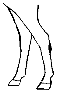
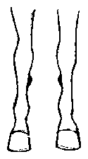
Body Conformation


Good leg conformation Bad leg conformation Bad leg conformation


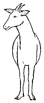


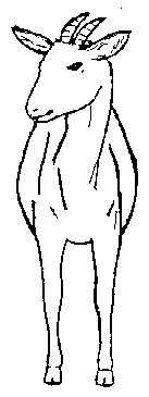


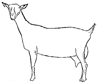


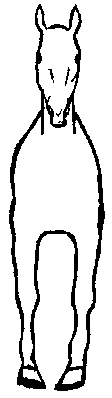

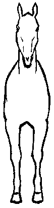


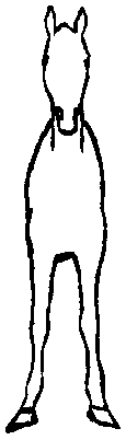

(Back legs too angled) (Back legs too straight)
Other faults commonly found in horses’ skeletal system: